Fig. 1.
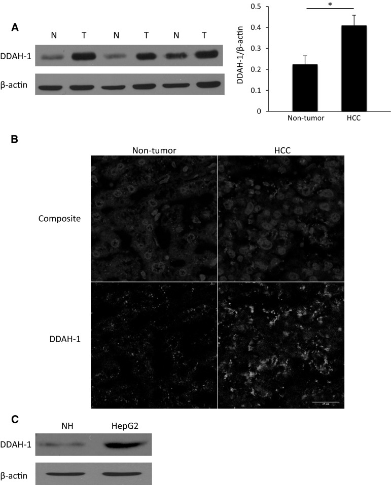
Overexpression of DDAH-1 in HCC. a DDAH-1 protein levels were measured with immunoblotting in paired HCC samples and their non-tumorous counterparts. Protein expression results were normalized to internal control β-actin. *P < 0.05. N non-tumorous liver (n = 20), T HCC tumor (n = 20). Imagings shown are representative results of three patients. b Non-tumorous human liver and human HCC tissues were stained for DDAH-1. In the composite images: Red, DDAH-1; blue, nuclei. In the single DDAH-1 channel images: DDAH-1, gray. c Expression of DDAH-1 in in vitro cultured human primary hepatocytes and in a HCC cell line was detected with immunoblotting analysis. (Color figure online)

