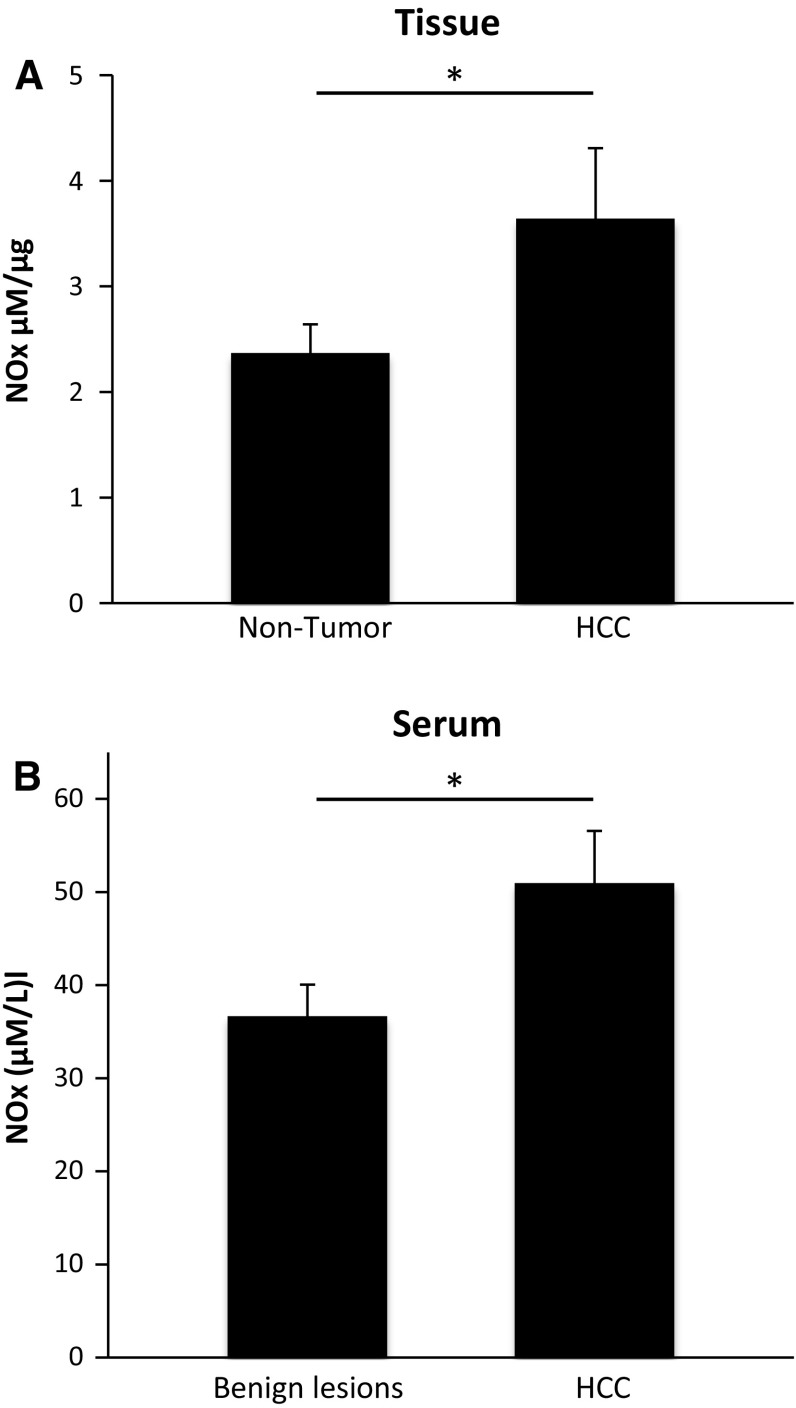
Fig. 3.
NO metabolites were increased in HCC tissue (n = 20) compared to non-tumorous liver tissue (n = 20) and were higher in serum of HCC patients (n = 20) compared to serum of patients with benign lesions (n = 10). NO metabolites were analyzed in tissue (a) and serum (b) by using a Griess reagent protocol. NOx concentration was defined as μM per μg protein in tissue homogenates and as μM per L serum. *P < 0.05

