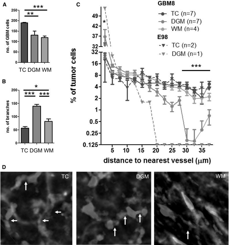
Fig. 5.

Topographical characteristics of GBM cells distant from the tumor core. a GBM cell density (per volume of 106 µm3) was calculated in the tumor core (TC, n = 7), and invasive fronts within the deep gray matter (DGM, n = 7) and white matter (WM, n = 4) of GBM8 tumors. b Number of vessel branches (per volume of 106 µm3 in the three aforementioned areas in GBM8 xenografted brains. c Proportion of GBM8 and E98 cells at indicated distance (µm) of the nearest vessel at distribution intervals of 2.5 µm. d GBM8 cells are interconnected via cell processes (arrows). *p < 0.05, **p < 0.01, ***p < 0.001, t test. Scale bars 10 µm. Samples were cleared with iDISCO. (Color figure online)
