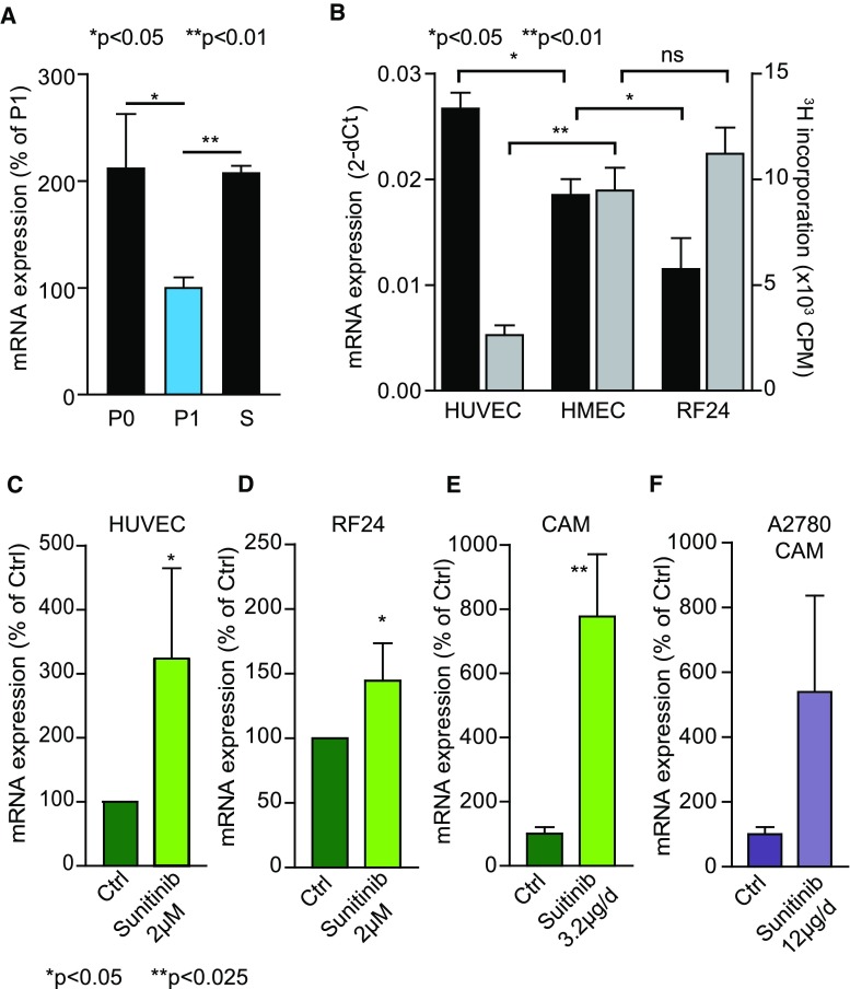
Fig. 3.
BRD7 expression is inversely related to endothelial cell growth. a HUVEC were isolated as described and either processed for RNA isolation immediately after isolation (P0) or grown under standard conditions for 3 days (P1), and BRD7 transcript levels were measured by qPCR. A clear reduction of BRD7 mRNA is evident in cells stimulated to proliferate in vitro. Serum withdrawal (S) of established HUVEC increases the BRD7 mRNA levels to that of primary isolates. *p < 0.05, **p < 0.01 t test. b BRD7 expression was measured in routinely cultured HUVEC, HMEC and RF24 by qPCR. In parallel, proliferation rate of the cells was measured by 3H-thymidine incorporation. Primary cells (HUVEC) show higher expression levels (black bars; left y-axis) than immortalized EC (RF24 and HMEC), but the opposite holds true for proliferation rate (gray bars; right y-axis). *p < 0.05 Wilcoxon test. c, d HUVEC (c) and RF24 (d) were treated with 2 µM sunitinib and BRD7 was quantified by qPCR. Sunitinib markedly increases BRD7 expression. *p < 0.05 Wilcoxon test. e CAMs were treated with 3.2 µg sunitinib per day, and chicken BRD7 expression was determined with qPCR. Similar to in vitro treatment, in vivo treatment with sunitinib increases BRD7 expression. **p < 0.01 ANOVA. f sunitinib treatment (12 µg/day) of A2780 tumors on the CAM results in an increase in vascular BRD7 expression as determined by qPCR. *p = 0.05 Mann–Whitney test. All data are presented as mean ± SEM

