Figure 5.
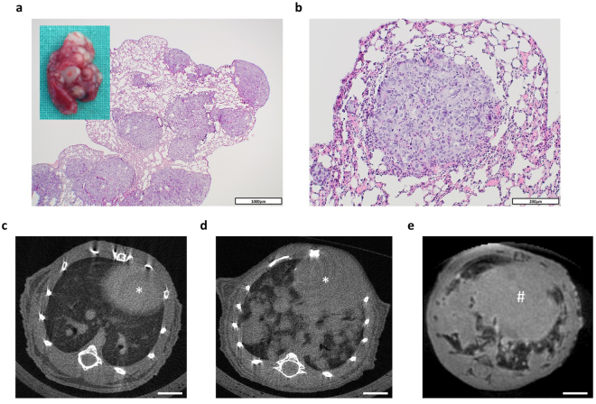
Development and detection of pulmonary metastases. (a) Representative microphotograph and macroscopic appearance (insert) of a lung bearing multiple metastases. Scale bar = 1000 μm. (b) Representative microphotograph of a single pulmonary metastasis. Scale bar = 200 μm. (c,d) Representative cross-sectional μCT images of a healthy lung (c) and a lung with multiple metastases (d). The heart is indicated by an asterisk (*). (e) Representative cross-sectional MRI-image (UTE, ultrashort echo time) of the basal parts of a lung with multiple metastases. The diaphragm is indicated by a rhomb (#). Scale bars = 3 mm.

