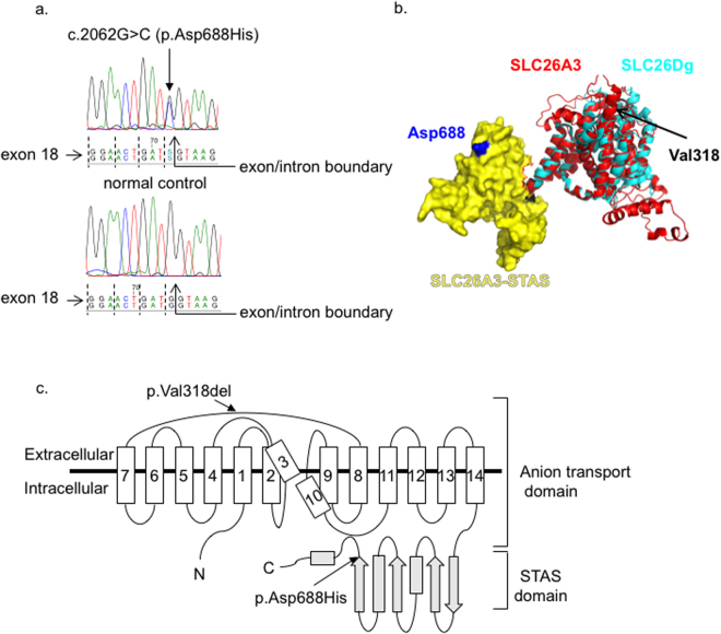
Figure 1.
Localization of the c.2062 G > C (p.Asp688His) variant. (A) The c.2062 G > C (p.Asp688His) variant is located at the last nucleotide position of the exon 18 of the SLC26A3 gene. Dashed lines indicate the reading frame of exon 18. (B) Putative 3D structure of the human SLC26A3. The transmembrane domain (ribbon, red) and the STAS domain (surface, yellow) representations of the human SLC26A3 (encompassing residues 1–699 of 764) were predicted based on their homology to the SLC26Dg structure (PDB_ID: 5DA0) (ribbon, cyan)43 by using Robetta online software (confidence 0.507600)67. The final model was generated using PyMol software (Schrödinger, Cambridge, MA). (C) The predicted 2D topology of the human SLC26A3 protein. The full-length protein (764 amino acids, AA) comprises 14 putative transmembrane domains—which form the ion transport domain—and the cytoplasmic C-terminal STAS domain. Arrows indicate the approximate positions of the Finnish founder mutation for CLD (c.949_951delGTG, p.Val318del) and for the c.2062 G > C (p.Asp688His) variant43. Note: In both the Slc26Dg crystal structure and our putative SLC26A3 model (B), the STAS domain orientation in respect to the transmembrane domain (TMD) suggests that the STAS domain is located within the phospholipid bilayer. Hence, this does not represent a native orientation since the STAS domain is expected to be found in the cytoplasmic C-terminal part of the protein.

