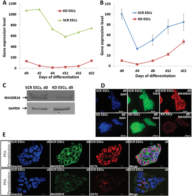
Figure 1.
Mageb16 expression in differentiated KD and SCR ESCs. (A) Mageb16 expression in undifferentiated and differentiated KD and SCR ESCs (microarray data). (B) qPCR analysis of Mageb16 expression in undifferentiated and differentiated KD and SCR ESCs (2 to 22 day EBs). (C) MAGEB16 protein expression in the KD and SCR ESCs. After preparation of the protein lysates, 10 µg protein was analysed by western blotting. Chemiluminescence detection of MAGEB16 has been performed using MAGEB16 polyclonal antibodies (1:250) and GAPDH has been detected using the anti-GAPDH antibody (1:2500) dilutions. (D) Cellular localization of MAGEB16 and SSEA1 in SCR and KD ESCs. Immunocytochemistry has been performed using primary anti-SSEA1 andibodies (1:50) (green colour) and anti-MAGEB16 antibodies (1:50) (red colour) and goat anti mouse IgM-alexa fluor 488 secondary antibody (1:1000) and goat anti rabbit IgG-alexa fluor 568 as secondary antibody (1: 1000). Cells were co-stained with nuclear marker Hoechst 33342. The overlay of nuclear and MAGEB16 staining reveals that the presence of MAGEB16 is restricted to cytosol and/or surface membrane (scale bar: 100 µm). (E) Confocal microscopy (upper panel+: permebilized cells; lower panel−: non-permebilized). Immunocytochemistry was performed using primary anti-OCT4 antibodies (1:250) (red color) and anti-MAGEB16 antibodies (1:50) (green color) and goat anti rabbit IgM-alexa fluor 488 secondary antibody (1:1000) and goat anti mouse IgG-alexa fluor 568 as secondary antibody (1:1000). Cells were co-stained with nuclear marker Hoechst 33342. The overlay of nuclear, OCT4 and MAGEB16 staining reveals the presence of MAGEB16, which is restricted to cytoplasmic domains of the ESCs (scale bar: 10 µm).

