Figure 5.
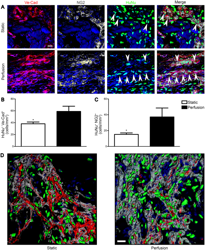
Perfusion-cultured human cells were directly involved in the formation of blood vessels. Patches were analysed after 28 days in vivo. (A) Representative immunofluorescence images of EC (Ve-Cad; red), pericytes (NG2; grey) and HuNu (green). White arrows indicate human cells directly involved in the formation of blood vessel Ve-Cad+ or NG2+. Quantification of the human cells Ve-Cad+ (B) and NG2+ (C) normalized over the analyzed area (n donor = 3). (D) 3D reconstruction of immunofluorescence images of EC (Ve-Cad; red), basal lamina (Laminin; grey) and HuNu (green). Nuclei were stained with DAPI (blue in A,D). *p < 0.05. Scale bar = 20 μm.

