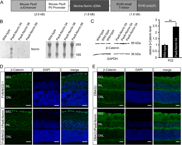
Figure 1.
Generation of transgenic Pax6-Norrin mice. (A) Schematic drawing of the transgene. (B) Northern blot analysis of Norrin mRNA in RNA of retinae from three transgenic lines (Pax6-Norrin-04, -06 and -69) and wild-type littermates (WT) at P2. Integrity of loaded RNA was controlled by methylene blue staining. (C) Western blot analysis for β-catenin in retinae from transgenic line Pax6-Norrin-04 and wild-type littermates (WT) at P22. β-catenin levels were analyzed by densitometry, normalized to GAPDH and plotted as x-fold to levels of wild-type controls (mean ± SEM; n = 9; **p < 0.01). (D,E) Immunostaining for β-catenin (green) in P22 retinae of wild-type and transgenic Pax6-Norrin-04 mice in the FVB/N/CD-1 background (D) and crossed with DBA/2J mice (E). Nuclei are labeled with DAPI (blue). GCL. ganglion cell layer; INL. inner nuclear layer; ONL. outer nuclear layer; Scale bars: 20 μm.

