FIG 8.
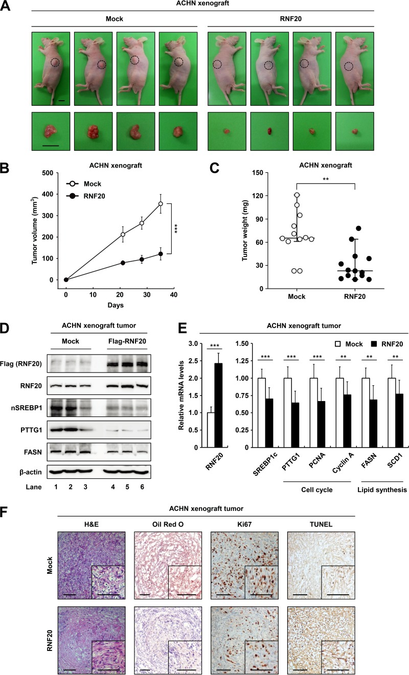
RNF20 overexpression attenuates tumor growth in xenograft mice. (A) Subcutaneous tumors of ACHN cells expressing either the negative control (Mock) or ectopic RNF20 were generated in female BALB/c nude mice. Representative images of tumors dissected at the end of the study showing the effect of RNF20 overexpression on the growth of xenograft tumors in vivo. Bar, 10 mm. (B) Xenograft tumor volumes (in cubic millimeters) of ACHN cells with or without ectopic RNF20 expression were determined over 35 days. The graph shows the means ± SEM; n = 10 for each group. (C) Endpoint xenograft tumor weights were determined and plotted. Data are represented as the means ± SEM; n = 10 for each group. (D) Expression levels of RNF20, nuclear SREBP1, PTTG1, and FASN protein in ACHN xenograft tumors were monitored using Western blotting. (E) The effects of ectopic RNF20 expression on cell cycle and lipogenic gene expression in ACHN xenograft tumors were determined using qRT-PCR. Relative mRNA levels are shown relative to the control group (Mock) levels. Data are presented as the means ± SD; n = 10 for each group; **, P < 0.01; ***, P < 0.001. (F) Histological analysis of xenograft tumors. Representative hematoxylin and eosin (H&E)- and Oil Red O-stained sections of negative-control (Mock)- or RNF20-transduced ACHN xenograft tumors. IHC of xenograft tumors stained with Ki67 and TUNEL. Bars, 100 μm.

