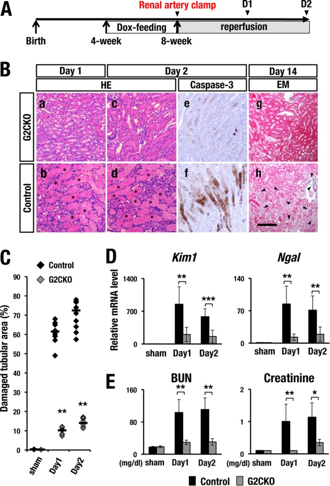
FIG 1.

G2CKO mice are resistant to renal IRI. (A) Experimental time course of Dox feeding and IRI induction. Transient renal artery clamping was performed at 8 weeks of age. The mice were subjected to analysis 1 day, 2 days, or 14 days after IR. (B, a to d) H&E staining of the kidney at 1 day and 2 days after unilateral renal IR. Note that control mice showed robust coagulative necrotic lesions in the cortical and outer medullary regions (b and d, asterisks), while G2CKO mice showed lower levels of necrosis (a and c). (e and f) Immunohistochemical analyses with an anti-cleaved-caspase-3 antibody. Note that control mice (f) showed a number of anti-cleaved-caspase-3-positive apoptotic cells in the proximal tubules 2 days after the IR. In contrast, G2CKO mice (e) showed very few apoptotic cells in the IRI kidney. (g and h) Elastica-Masson (EM) staining of IRI kidneys from G2CKO (g) and control (h) mice 14 days after unilateral IR (day 14). Control mouse kidneys showed robust tubular injury, while G2CKO kidneys showed well-preserved renal tubules. Scale bar, 200 μm. (C) Quantitative analysis of damaged tubular area in H&E-stained kidney sections. Percentages of damaged tubular area were significantly decreased in the G2CKO kidneys 1 and 2 days after the unilateral IR compared with control mouse kidneys. The data points represent individual values from each section. The bars indicate the median of each group. (D and E) The expression levels of tubular injury biomarkers (D), as well as BUN and serum creatinine levels (E), 1 and 2 days after IR and 2 days after the sham operation in the kidneys of G2CKO and control mice. Note that the levels of tubular injury biomarkers, BUN, and serum creatinine were maintained at a lower level after IRI in G2CKO mice than in control mice. The data are presented as means ± SD (n = 5 in each group of mice). The statistical significance is indicated (***, P < 0.005; **, P < 0.01; *, P < 0.05).
