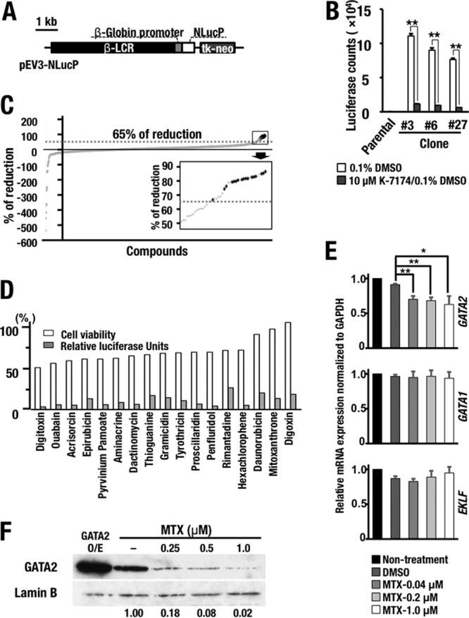
FIG 5.

High-throughput screening identifying GATA factor inhibitors. (A) Schematic diagram of the NanoLuc (NLucP) reporter construct. (B) Luciferase activity in the K562-NLucP reporter cell lines. Four thousand cells of three K562-NLucP cell lines (number 3, number 6, and number 27) carrying the NanoLuc reporter construct were exposed to 0.1% DMSO solvent or 10 μM Κ-7174 for 24 h. The results are shown as means and SD, with the experiments performed in triplicate (**, P < 0.01). Parental K562 cells served as a control. (C) Primary chemical library screening data for 1,165 compounds in the off-patent drug library of Keio University. The data are presented as the percent reduction relative to the value obtained for the 0.1% DMSO-treated control. The inset, a magnified view of the boxed area on the upper right, shows the top 29 compounds, which were subjected to the second screening. The dotted lines indicate the 65% reduction level. The 17 hit compounds are indicated by black dots. (D) Validation of the hit compounds. Shown are the percent luciferase activity and cell viability compared with the value for the 0.1% DMSO-treated control after 28-hour exposure to 10 μM each compound. (E) Expression levels of GATA1, GATA2, and EKLF mRNAs in K562 cells treated with MTX. Note that MTX treatment predominantly diminished the GATA2 mRNA level, while the treatment did not affect the GATA1 and EKLF mRNA levels in K562 cells. The statistical significance is indicated (**, P < 0.01; *, P < 0.05). (F) MTX treatment diminishes the GATA2 protein expression level in HEK293T cells in a dose-dependent manner. The far left lane contained HEK293T cells overexpressing (O/E) GATA2 employed as a control.
