
FIG 1.
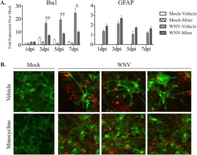
Minocycline treatment inhibits microglial activation during WNV infection of SCSC. (A) RT-qPCR analyses were performed for Iba1 and GFAP expression in SCSC taken at 1 dpi, 3 dpi, 5 dpi, and 7 dpi. Changes in gene expression levels are indicated as fold increase over mock infection, with 1-dpi mock-infected, vehicle-treated SCSC used as the normalized control sample (expression set as 1) and beta-actin used as the normalized control gene. Iba1 gene expression (left) increased in WNV-infected (gray bars) compared to mock-infected (open bars), vehicle-treated SCSC in a time-dependent manner, indicating microglial activation. Minocycline (Mino) treatment of WNV-infected SCSC (hatched gray bars) caused significant decreases in Iba1 expression compared to WNV-infected, vehicle-treated counterparts (gray bars). GFAP gene expression (right) increased in WNV-infected, vehicle treated SCSC (gray bars) compared to mock-infected, vehicle-treated controls (open bars), but not as dramatically nor as high as Iba1. There was no decrease in WNV-induced GFAP expression following minocycline treatment. The asterisks indicate statistically significant differences in Iba1 expression in minocycline-treated versus vehicle-treated, WNV-infected SCSC (*, P < 0.05; **, P < 0.01; unpaired Student t test). The error bars indicate standard errors of the mean. Fifty SCSC (n = 2 mice) were used per experimental condition. (B) Immunohistochemistry of Iba1 (green) and WNV-E (red), imaged with a 60× objective, from mock-infected (top row) and WNV-infected (bottom row) SCSC at 6 dpi. Iba1 expression was increased in WNV-infected SCSC compared to mock-infected samples, but the ameboid and enlarged microglial cells seen in vehicle-treated SCSC were absent from minocycline-treated SCSC.