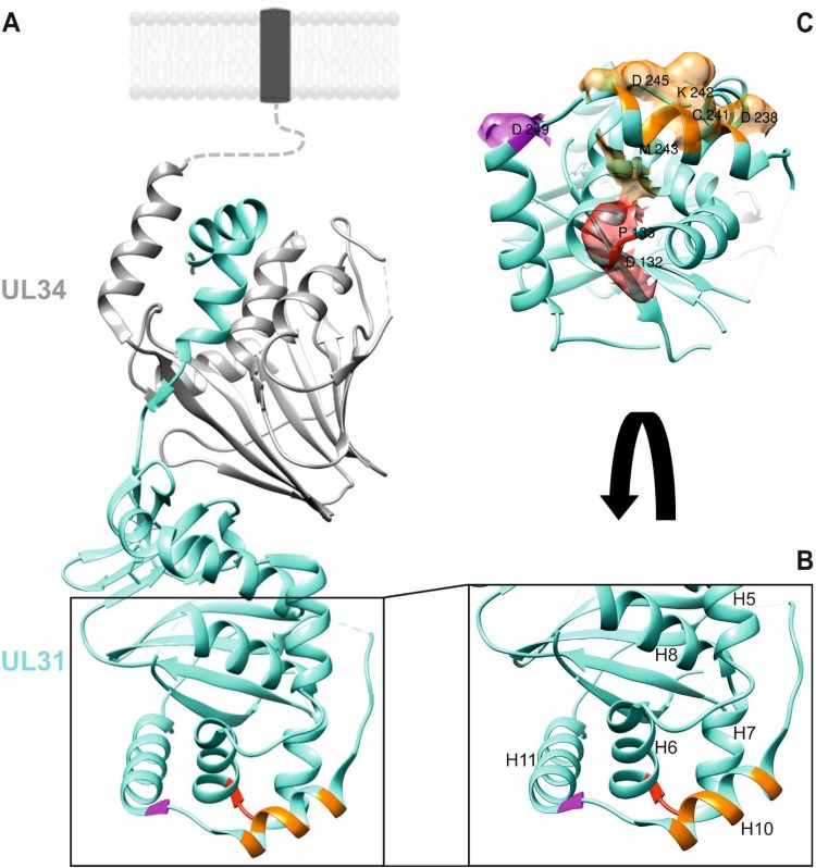
FIG 1.
Location of putative capsid interaction interfaces in PrV pUL31. (A) In the PrV NEC structure (19), the pUL34 component is shown in gray, and pUL31 is shown in turquoise. Orientation toward and anchorage in the inner nuclear membrane or the primary virion envelope is indicated by the dotted line, and the transmembrane anchor is represented by a dark box. Location of amino acids mutated in this study is indicated in red (D132 and P133), orange (D238, C241, K242, M243, and D245), and magenta (D249). (B) Enlarged membrane-distal end of the NEC in ribbon presentation with alpha-helices numbered, as described previously (19). (C) Amino acids changed in this study are shown in surface presentation using the same coloring. Molecular graphics and analyses were performed with the UCSF Chimera package (35).

