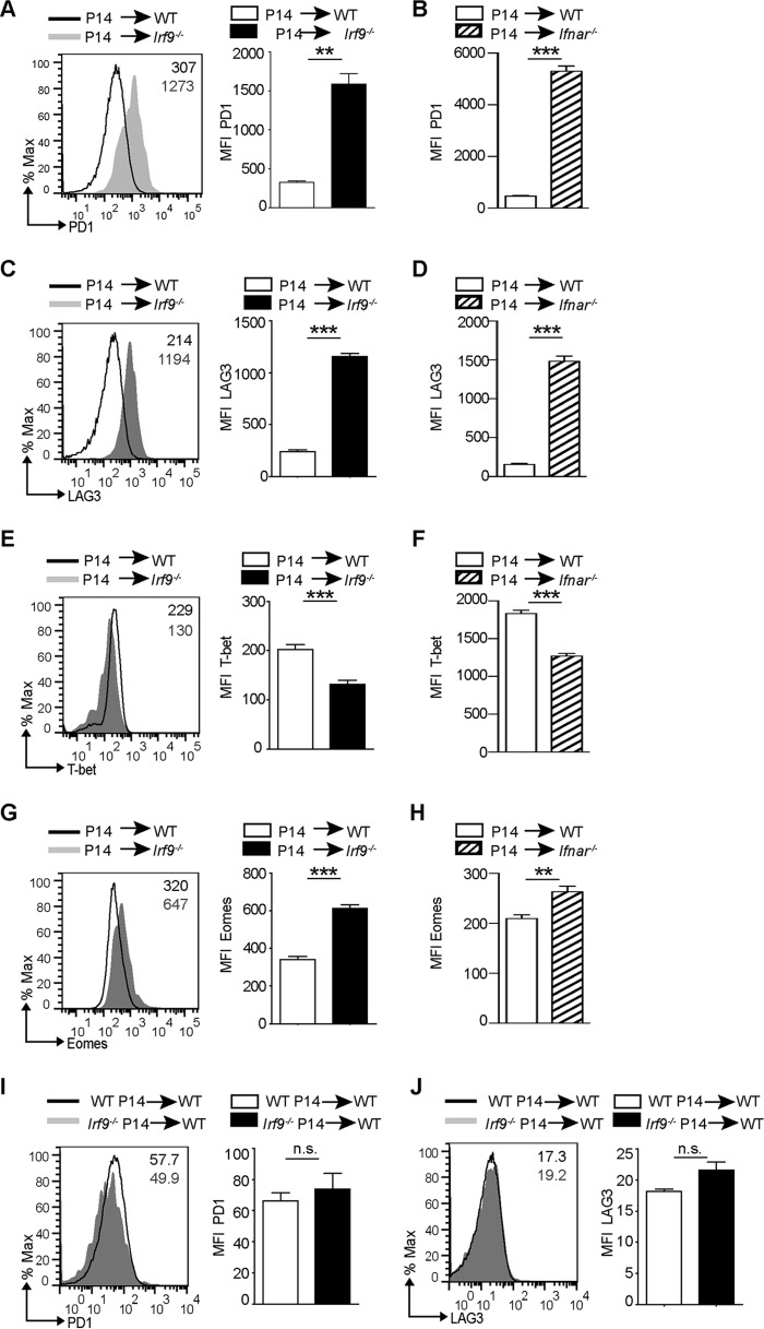
FIG 4.
IRF9 extrinsically regulates exhaustion of CD8+ T cells. Prior to LCMV-Arm infection, 104 negatively sorted CD8+ T cells from CD45.1+ P14 mouse cells were transferred into WT or Irf9−/− mice (A, C, E, and G) or into WT or Ifnar−/− mice (B, D, F, and H). Spleens were analyzed at day 8 p.i. (A to H) MFI for LAG3, PD1, Eomes, and T-bet of P14 cells transferred into WT, Irf9−/−, or Ifnar−/− mice. Representative histograms are shown. Bar diagrams display means and SEM (n = 5 per group). (I and J) Prior to LCMV-Arm infection, 104 negatively sorted CD8+ T cells from CD45.2+ P14 mice or from CD45.2+ Irf9−/− P14 mice were transferred into CD45.1+ WT mice. The spleens were analyzed at day 8 p.i., and the graphs show MFI for PD1 and LAG3 of WT or Irf9−/− P14 cells transferred into WT mice. Representative histograms are shown. Bar diagrams to the right display means and SEM (n = 5 per group). For panels A to F, data from one of two independent experiments with consistent results are shown. **, P < 0.01; ***, P < 0.001; n.s., not significant (unpaired two-tailed Student's t test).

