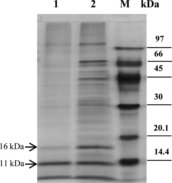
FIG 4.

Protein pattern of MetSV. Total protein extracts of MetSV were separated by SDS-PAGE, followed by silver staining. Lane 1, MetSV protein extract after a second washing step of the virus particles; lane 2, MetSV protein extract without a second washing step; M, low-molecular-weight marker (GE Healthcare Europe GmbH, Freiburg, Germany).
