FIG 4.
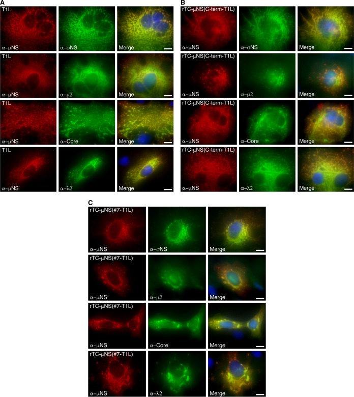
Colocalization of MRV proteins with recombinant virus-expressed μNS. CV-1 cells were infected with T1L (A), rTC-μNS(C-term-T1L)/P2 (B), or rTC-μNS(#7-T1L)/P2 (C) and at 18 h p.i. were immunostained with mouse (second and third rows) or rabbit (first and fourth rows) antibodies against μNS and mouse σNS antibodies (first row), rabbit μ2 antibodies (second row), MRV core rabbit antibodies (third row), λ2 mouse antibodies (fourth row), followed by Alexa 594-conjugated donkey α-rabbit or α-mouse IgG (first column) and Alexa 488-conjugated donkey α-mouse or α-rabbit IgG (second column). Merged images with DAPI-stained nuclei are also shown (third column). Bars, 10 μm.

