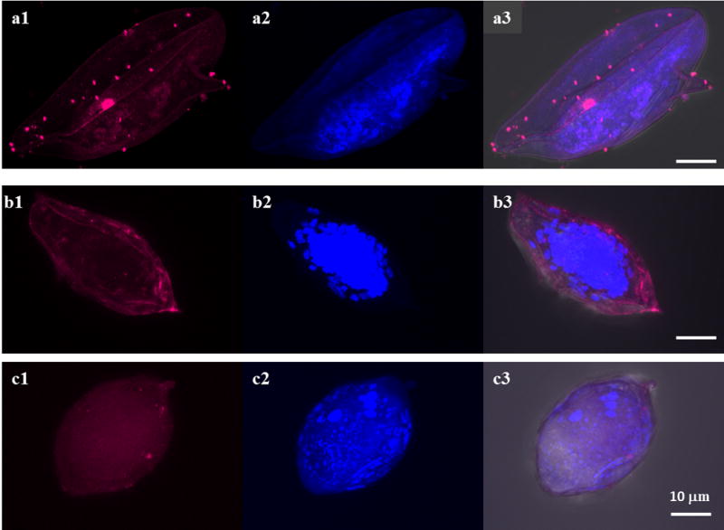
Fig. 1.

Localization of Sm-p80 in Schistosoma mansoni, S. haematobium and S. japonicum eggs. Representative fluorescence images of S. mansoni, S. haematobium and S. japonicum eggs respectively are shown in panels a, b and c. The distribution of Sm-p80/Sm-p80 orthologs is shown in a1, b1 and c1 images (magenta). The eggs were labeled with the rabbit antibody against Sm-p80. a2, b2 and c2 show DAPI labeling which stains nuclei (blue). The samples were mounted using Prolong Gold antifade reagent with DAPI. a3, b3 and c3 show an overlay showing the distribution of sm-p80/Sm-p80 orthologs (magenta) and DAPI (blue) in differential interference contrast (DIC). The images were taken using a Nikon T1-E confocal microscopy with a 60× objective and analyzed with NIS software. The eggs images represent a maximum projection intensity derived from a Z-stack. Scale bars, 10 μm
