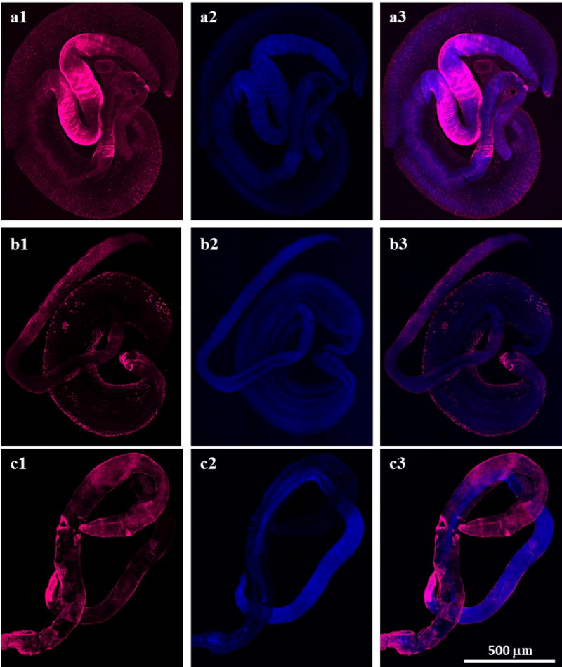
Fig. 4.

Sm-p80/Sm-p80 ortholog protein is localized on the tegument of Schistosoma mansoni, S. haematobium and S. japonicum adult worms. Representative stitched fluorescence images of S. mansoni, S. haematobium and S. japonicum adult worms respectively are shown in panels a, b and c. a1, b1 and c1 show the distribution of sm-p80 and Sm-p80 ortholog proteins. The S. mansoni adult worms were labeled with the rabbit antibody against Sm-p80 (a1), the S. haematobium with the mice serum against Sm-p80 ortholog protein (b1) and the S. japonicum with the baboon serum against Sm-p80 ortholog protein (c1). The samples were mounted using Prolong Gold antifade reagent with DAPI. a2, b2 and c2 show DAPI labeling which stains nuclei (blue). a3, b3 and c3 show an overlay of the distribution of sm-p80/Sm-p80 ortholog proteins (magenta) and DAPI (blue). The images taken using a Nikon T1-E confocal microscopy with a 10x objective were stitched and analyzed with NIS software. All images represent a maximum projection intensity derived from a Z-stack. Scale bars, 500 μm
