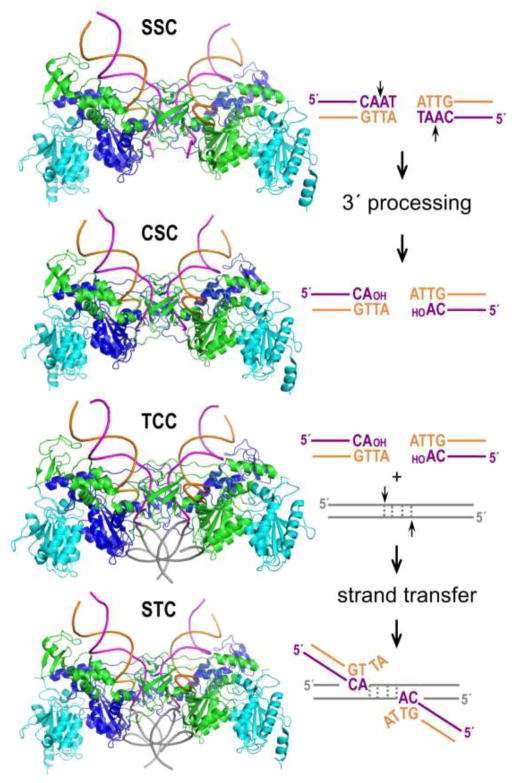
Figure 1.
Retroviral integration and intasome nucleoprotein complexes. Shown are representative PFV intasome structures (SSC, pdb accession code 4e7h; CSC, pdb code 3oy9; TCC, pdb code 3os2; STC, pdb code 3os0). The intasomes consist of purified recombinant IN protein and synthetic oligonucleotides that model the U5 ends of viral DNA. Two DNA binding IN protomers are painted blue and green, whereas supportive IN molecules are cyan. Adjacent DNA end sequences are color-coded to match the transferred magenta and non-transferred orange viral DNA strands in the structures. Short vertical arrows, scissile phosphodiester bonds. Target DNA is shown in grey; during strand transfer, the DNA is cleaved with a 4 bp staggered cut.

