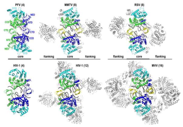
Figure 2.
Retroviral intasome structures and the CIC. The PFV structure is an underside view of the CSC relative to Figure 1, labeled for individual IN domains. The other intasome structures (MMTV CSC, pdb code 3jca; RSV STC, pdb code 5ejk; tetrameric HIV-1 STC, pdb code 5u1c; MVV CSC; pdb code 5m0q) are similarly oriented, size matched for common CIC components (colored). Synaptic CTDs that are donated from flanking IN dimers are shown in yellow. The portions of these structures that do not form part of the CIC are deemphasized by light gray. Viral DNA strands are black, and the target DNA strands from the RSV and HIV-1 STC structures, as well as the LEDGF/p75 IN-binding domain from the HIV-1 dodecamer, were omitted for clarity. Numbers indicate the number of IN molecules within each structure.

