Figure 2.
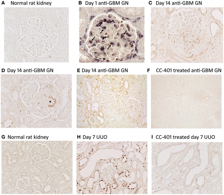
JNK activation in kidney injury. Detection of JNK activation by immunohistochemistry staining for c-Jun phosphorylated at Ser63 (p-c-Jun). (A–F) JNK activation in rat anti-glomerular basement membrane (GBM) glomerulonephritis (GN). (A) Normal rat glomeruli lack p-c-Jun staining. (B) Two color immunostaining for p-c-Jun (brown nuclei) and ED1+ macrophages (purple/blue) in a glomerulus on day 1 of anti-GBM disease. Arrows indicate cells stained for both ED1 and p-c-Jun, demonstrating JNK signaling in macrophages. Arrowhead shows p-c-Jun immunostaining in a podocyte-like cell. Parietal epithelial cells staining for p-c-Jun is also evident. (C) Glomerulus on day 24 of anti-GBM disease shows nuclear p-c-Jun staining of cells in the capillary tuft and in the crescent. (D) Day 24 of anti-GBM disease showing p-c-Jun staining in a multinucleated giant cell in a glomerular crescent (*). (E) Lower power view of day 24 anti-GBM disease showing p-c-Jun+ cells in the glomerular tuft, in crescents, in many tubular epithelial cells and in some interstitial cells. (F) Treatment with the JNK inhibitor CC-401 over days 7–24 of anti-GBM disease blocks c-Jun phosphorylation. (G–I) JNK activation in the rat unilateral ureteric obstruction (UUO) model. (G) Normal rat kidney shows a lack of p-c-Jun staining in glomeruli and tubules. (H) Day 7 after UUO shows extensive JNK activation in both atrophic tubules and in tubules with normal morphology. Glomeruli do not show inflammation in this model and they lack p-c-Jun staining. (I) Treatment with the JNK inhibitor CC-401 over days 0–7 of the UUO model blocks c-Jun phosphorylation. Reproduced with permission from Flanc et al. (2007) (A,B); Ma et al. (2009) (C–F), and; Ma et al. (2007a) (G–I).

