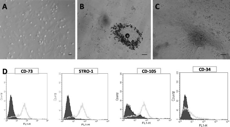
Fig. 1.
In vitro culture and characterization of caprine BM-MSCs. A Morphology of caprine bone marrow derived mesenchymal stem cells exhibiting typical fibroblastoid phenotype in primary culture (Scale bar 30 μm). B Adipogenesis induced lipid droplets observed in red colour after specific Oil Red O staining in in vitro expanded MSCs (Scale bar 50 μm). C Osteogenic differentiation of MSCs, brownish black coloured mineral deposition as demonstrated by von Kossa staining (Scale bar 50 μm). D Flowcytometric analysis of surface antigens in in vitro expanded caprine Bone marrow derived MSCs. Cells were stained with primary antibodies directed against CD-73, STRO-1, CD-105, CD-34 and counter stained by FITC conjugated secondary antibodies. Calibrated histogram represents the number of events on the Y-axis and FITC-fluorescent intensity (FLH-1) on X-axis. The shadowed histogram indicate negative controls. (Color figure online)

