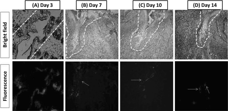
Fig. 3.
Persistence of PKH26 labelled caprine BMMSCs at different time points post transplantation when observed under bright field and fluorescent microscopy. Dotted line represents the site of injury. White arrows points the distribution of PKH26 labelled cells segregated along the wound margin and within wound bed. (Color figure online)

