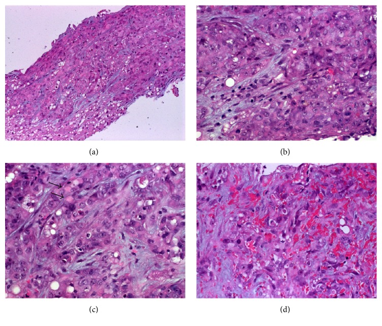
Figure 2.
(a) Liver biopsy showed tumor tissue diffusely infiltrating the liver parenchyma (normal liver is seen in lower left field). (b) Higher magnification showed anaplastic large epithelioid cells with prominent nucleoli. (c) Scattered rhabdoid inclusions are seen (arrows) as well as small cytoplasmic vacuoles possibly suggestive of abortive glandular differentiation. (d) In other areas, hemorrhage and spindling might be mistaken for a vascular neoplasm.

