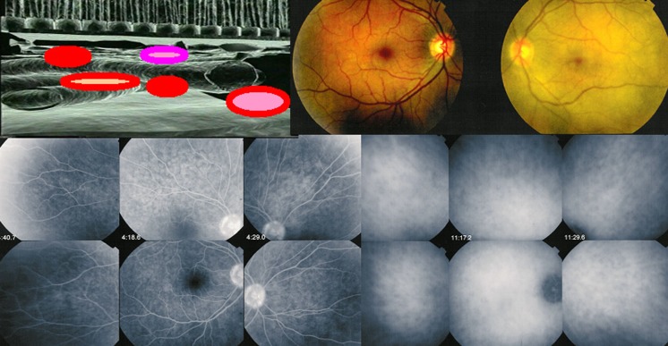
Fig. 1.
During the prodromal stage of the disease, subclinical choroidal inflammation is silently developing in the choroidal stroma as shown on cartoon (top left). This subclinical choroidal involvement can only be detected by ICGA, possibly by choroidal OCT. On the fundus pictures shown on top right, the right fundus is discoloured yellow due to massive choroidal infiltration. The right fundus looks normal and this patient was diagnosed as “unilateral” VKH disease. FA shows no lesions (six bottom left frames) but ICGA (six bottom right frames) clearly shows numerous hypofluorescent dark dots (HDDs) indicating choroidal granulomas in the apparently uninvolved eye

