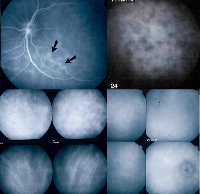
Fig. 4.
Indocyanine green angiographic signs. ICGA is the only technique to analyse choroidal inflammatory signs including early stromal hyperfluorescent vessels (top left), hypofluorescent dark dots (HDD) indicating choroidal granuloma (top right), fuzzy indistinct choroidal vessels (top 2 frames of bottom left quartett). After 3 days of intravenous 1000 mg daily methylprednisolone the normal pattern of vessels is again recognizable (bottom 2 frames of bottom left quartett). Bottom right quartett of frames shows diffuse late hyperfluorescence and hyperfluorescent inflamed disc

