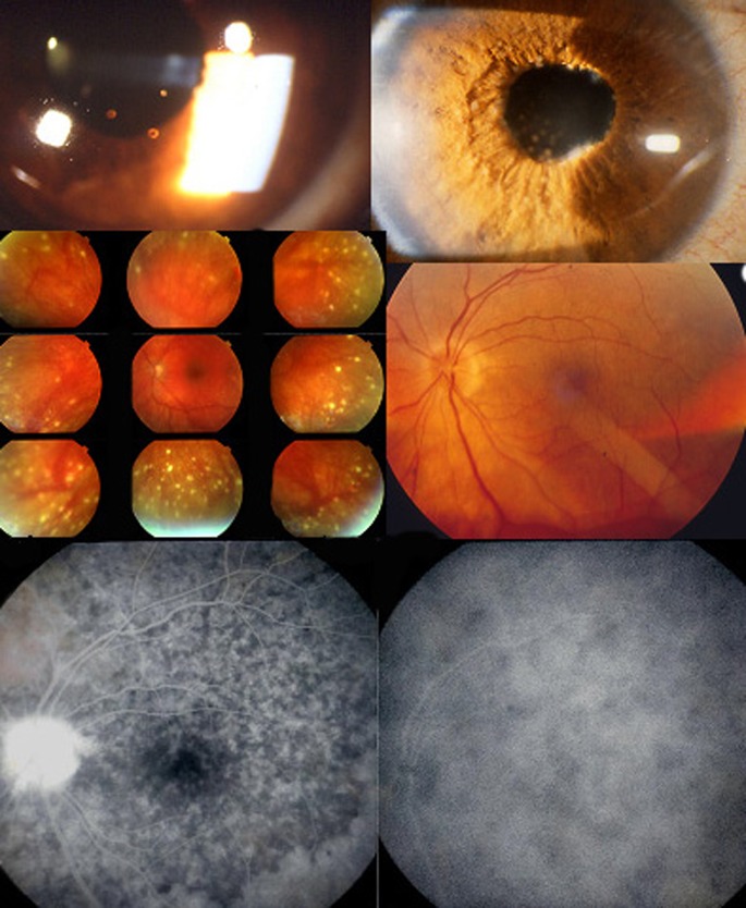
Fig. 6.
Signs found in chronically evolving disease. Chronic granulomatous uveitis with old pigmented KPs (top left), Koeppe nodules, iris infiltration and Busacca nodules (top right). Mid-periphery hypopigmented lesions are shown on the middle left picture and sunset-glow-fundus on the right middle picture. Mottled irregular, disturbed RPE in the posterior pole and high-water marks indicating limit of reattached serous retinal detachment as well as disc hyperfluorescence seen on the FA frame (bottom left). Only ICGA can show that disease is still active as evidenced by the numerous dark dots (bottom right)

