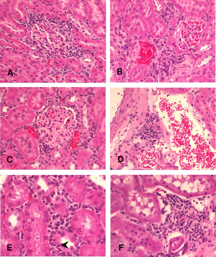
Fig. 3.
Histopathology of the low-dose (1.1 mg/m3) exposure group. Periglomerular inflammation at 24 hours, 7 days, and 30 days after exposure (A–C). Perivascular inflammation at 7 days after exposure (D). Interstitial inflammation with tubulitis featuring lymphocytic infiltrate within the tubular epithelium (arrowhead) at 24 hours after exposure (E). Interstitial inflammation at 30 days after exposure (F). Hematoxylin and eosin stain, original magnification = 200×.

