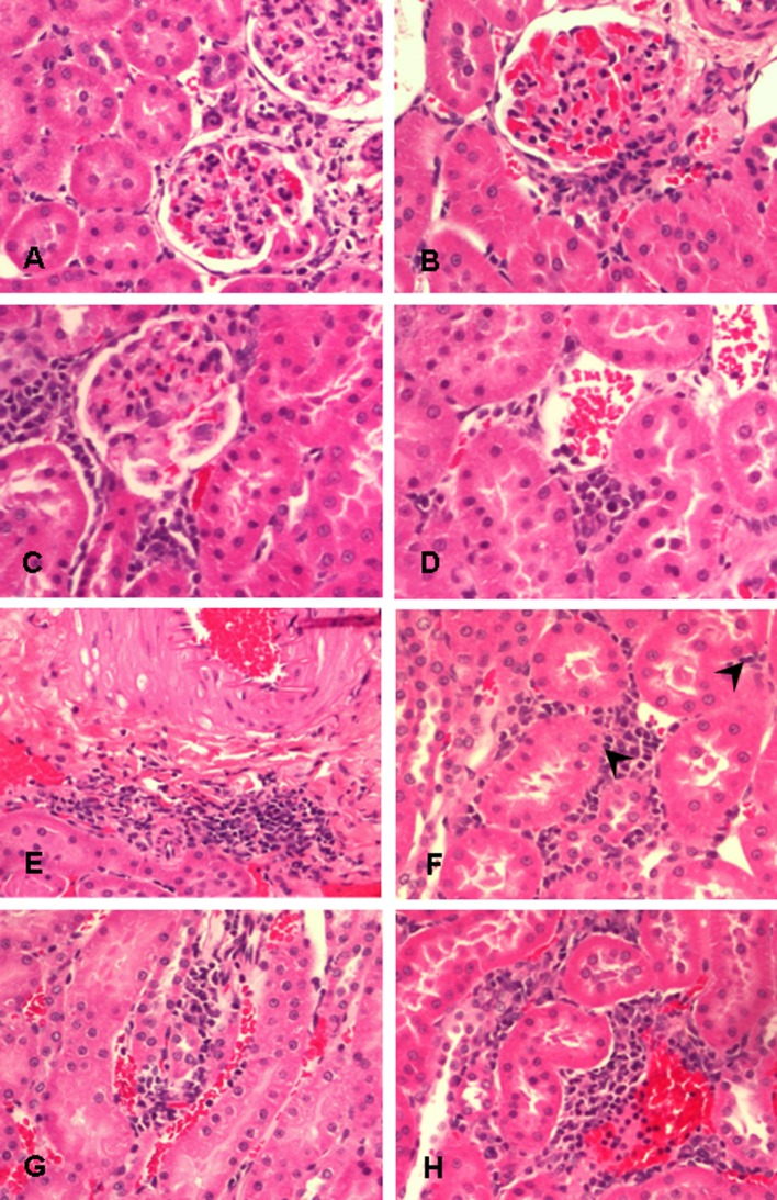
Fig. 4.
Histopathology of the high-dose (4.9 mg/m3) exposure group. Periglomerular inflammation at 24 hours, 7 days, and 30 days after exposure (A–C). Perivascular inflammation at 24 hours and 7 days after exposure (D, E). Interstitial inflammation with tubulitis featuring lymphocytic infiltrate within the tubular epithelium (arrowhead) at 24 hours after exposure (F). Interstitial inflammation at 7 days and 30 days after exposure (G, H). Hematoxylin and eosin stain, original magnification = 200×.

