
Figure 3.
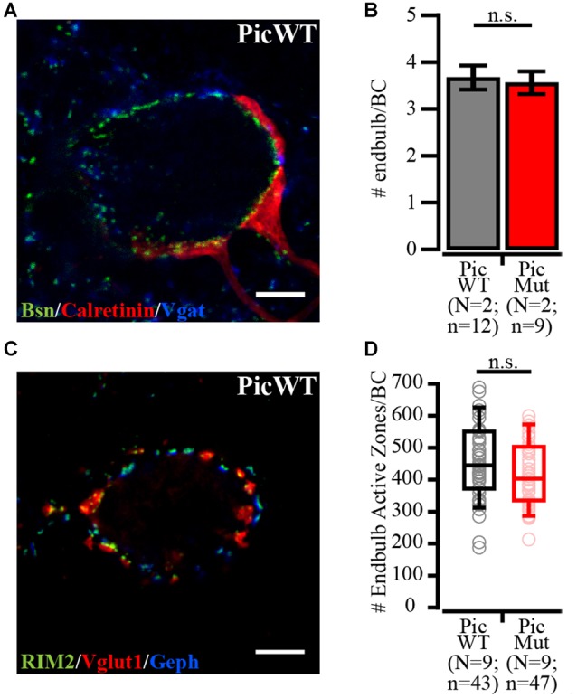
Number of endbulbs and endbulb AZs per bushy cell in aVCN. (A) Confocal section of a bushy cell in PicWT labeled with Bassoon (Bsn; AZ marker), Calretinin (endbulbs of Held) and Vgat (inhibitory presynaptic terminals). (B) Number of endbulbs converging onto a bushy cell was quantified by visually tracing and counting Calretinin-stained endbulbs. PicWT (N = 2; n = 12) and PicMut (N = 2; n = 9) receive comparable number of endbulbs (n.s. p-value ≥ 0.05, Student’s t-test). (C) Confocal section of a bushy cell in PicWT labeled with RIM2 (AZ marker), Vglut1 (excitatory synapses) and Gephyrin (Geph, inhibitory synapses). (D) Number of endbulb AZs (approximated from the # of excitatory AZs) per bushy cell quantified by subtracting the number of inhibitory AZs (AZ marker puncta juxtaposed with Gephyrin) from the total number of AZ marker puncta. Endbulb AZ number in PicWT (N = 9; n = 43) and PicMut (N = 9; n = 47) was comparable (n.s. p-value ≥ 0.05, Wilcoxon rank sum test). Data information: Box and whisker plot presents median, lower/upper quartiles and 10–90th percentiles. Bar plot represents mean ± SEM (error bars). N, number of animals; n, number of BCs.