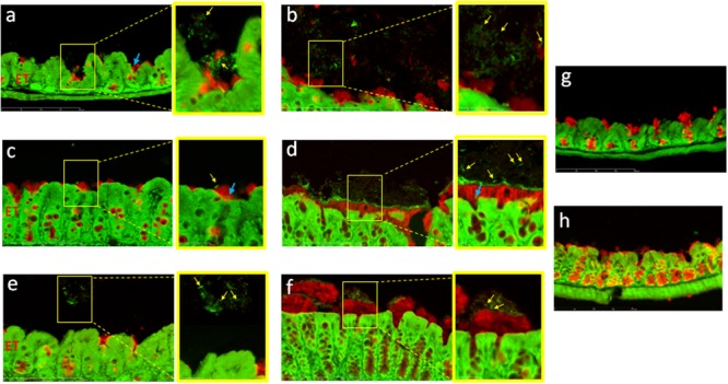
FIGURE 3.

Clostridium difficile cells are mainly localized at the surface of the mucus layer. Immunodetection of mucus and fluorescent labeling of bacteria by SYTO® 9 in the cecum (a,c,e,g) and in the colon (b,d,f,h) of mice infected with strains 630Δerm (a,b), R20291 (c,d), P30 (e,f), or in axenic mouse (g,h). Mucus is stained in red and DNA (bacterial, eukaryotic intracellular and, extracellular DNA) is stained in green. The right panel is the enlarged section of the yellow boxed portion of the image. Yellow arrows indicate position of bacteria outside the mucus layer. Blue arrows indicate position of bacteria in the outer layer of mucus. Scale bar is 200 μm.
