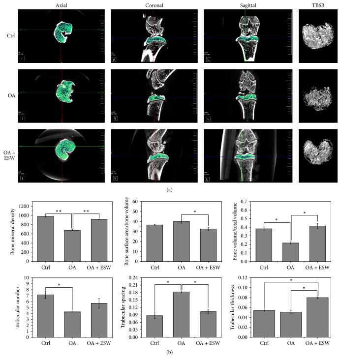
Figure 2.
Characterization of OA-induced subchondral bone changes in rats. (a) TBSB = three-dimensional reconstruction of trabecular bone of the subchondral bone. Micro-CT images of the joint tibial condyle subchondral bone at 8 weeks were different in the control versus ESW stimulation groups. Structural integrity of the subchondral bone was enhanced in the OA + ESW group compared with the OA group. (b) Micro-CT analysis of subchondral bone to evaluate knee joint function after ESW treatment in the MIA-induced OA rat model. BMD, BSA/BV, BV/TV, Tr.N, Tr.S, and Tr.Th data for all groups. ∗P < 0.05 and ∗∗P < 0.01 compared with control (error bar is SEM).

