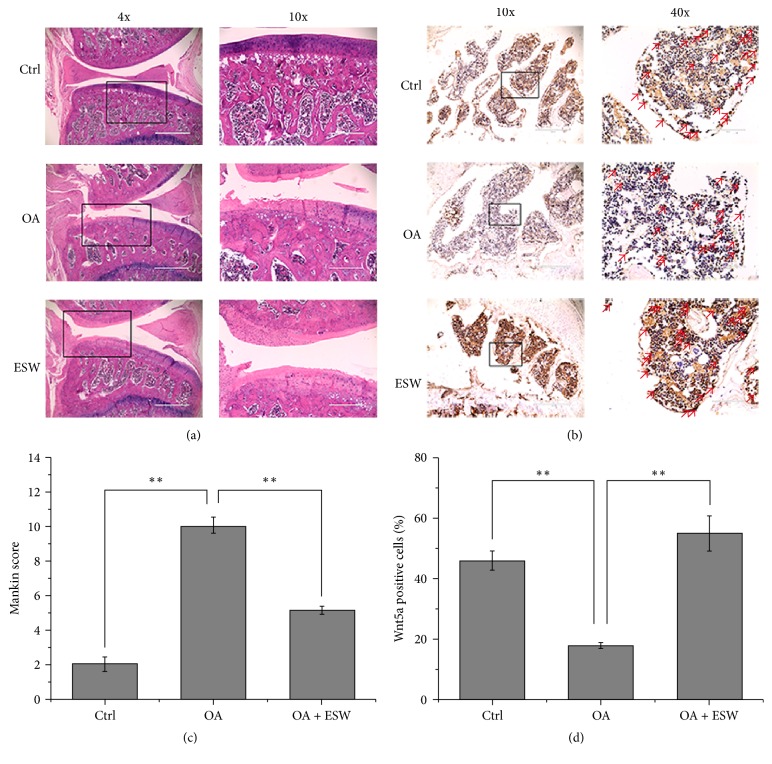
Figure 3.
Histopathologic assessment of cartilage and subchondral bone. (a) Hematoxylin-eosin (HE) staining of cartilage and subchondral bone at the 8th week after OA models were established successfully. (b) Wnt5a immunohistochemical (IHC) staining of subchondral bone of knee joint tibial condyle from 8-week control or experimental groups. The red arrows represent staining positive cells. (c) The OA group showed significant increases in Mankin score that was comparable to that of control. The ESW appeared effective in OA + ESW group compared with OA group (∗∗P < 0.05; error bar is SEM). (d) The number of Wnt5a-positive cells within the selected area was counted. Significant decreases in Wnt5a in the OA group compared with the control. The OA + ESW group showed significant increases in Wnt5a that were comparable to the OA (∗∗P < 0.05, error bar is SEM).

