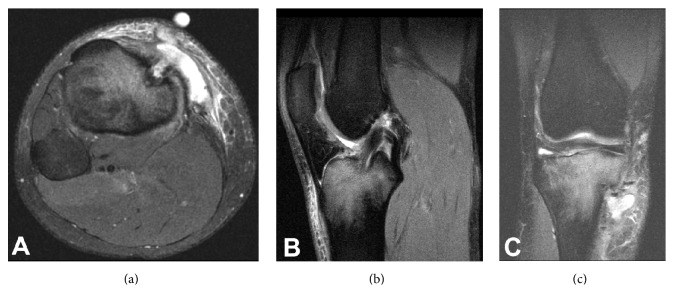
Figure 2.
(a) Axial slice of the MRI demonstrating the pretibial fluid collection in continuity with the orifice of the tibial tunnel. (b) T2 sagittal image demonstrates an intact ACL graft with adjacent tibial bone marrow edema. (c) T2 coronal image again demonstrates the pretibial fluid collection in continuity with the tibial tunnel and proximal tibial edema.

