
Figure 1.
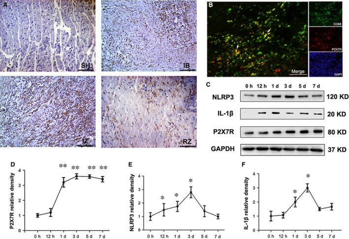
Expression profile of the P2X7R/NLRP3 inflammasome components after myocardial infarction (MI). (A) Immunohistochemical staining of P2X7R 3 days post‐MI in sham‐operated tissue (SH), the MI‐infarcted border (IB), the MI‐infarcted zone (IZ) and the remote zone (RZ). An increased expression of P2X7R was observed in the inflammatory cells and myocytes of infarcted tissue (IZ) and in the infarcted border (IB) at d3 post‐MI. (B) Double‐immunostaining for CD68 (green) and P2X7R (red) in the vehicle‐MI group showed a limited distribution of P2X7R on macrophages in the infarcted border. (C) Western blot showing the expression profiles of NLRP3 (120 kD), mature IL‐1β (17 kD) and P2X7R (80 kD) in the left ventricle 0 hr, 12 hrs, 1, 3, 5 and 7 days post‐MI. P2X7R, NLRP3 and mature IL‐1β were quantified relative to the GAPDH (37 kD) levels (D, E and F) (n = 5 per group and per time‐point). Bar = 30 μm. *P < 0.05 and **P < 0.01 versus 0 hr.