
Figure 1.
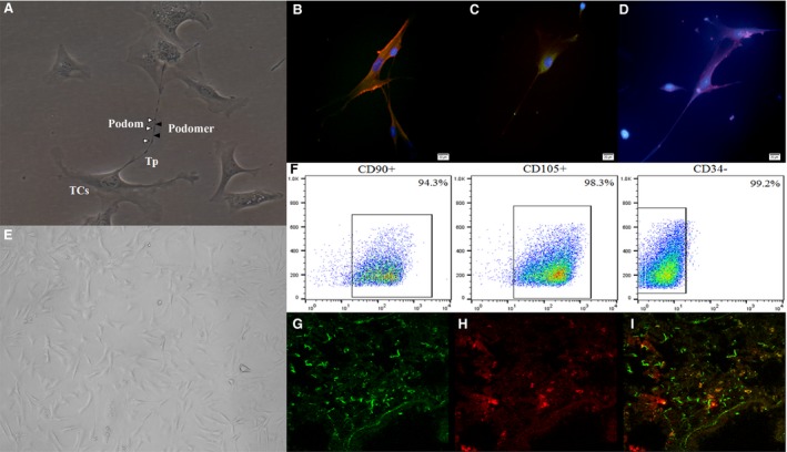
The identification and lung distribution of telocytes (TCs) and mesenchymal stem cells (MSCs). (A) The morphology of TCs under the light microscope. The bead‐like extending cellular process was named as telopode (Tp), with alternation of podoms (▵) and podomers (▲) (×200). (B) vimentin (Red) and CD34 (Green) fluorescence staining was positive. (C) CD34 (Green) fluorescence staining was positive. (D) c‐kit (Red) fluorescence staining was positive (×200). (E) The morphology of MSCs under the light microscope (×200). (F) The surface antigen CD90 and CD105 of MSCs were positive, and CD34 was negative. The mice lung tissue of TCs + MSC group, (G) CFSE‐labelled MSCs (Green); (H) PKH26‐labelled TCs (Red); (I) The merged image of two cells (n = 4/group).