
Figure 1.
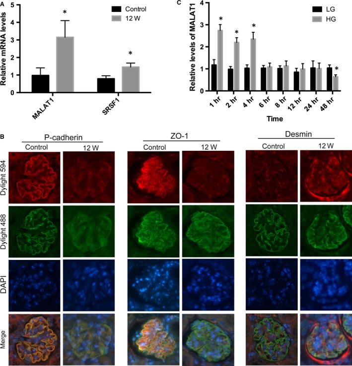
STZ‐induced diabetic podocyte injury and MALAT1 alterations in vivo and in vitro. (A) PCR analysis showed MALAT1 and SRSF1 were significantly up‐regulated in the kidney at 12 weeks after the onset of diabetes, in comparison with the control. Values denote the mean ± S.D.; *P < 0.01 versus control. (B) Immunofluorescence presented that in the control mice, p‐cadherin and ZO‐1 were intensely expressed in a leaner setting along the glomeruli, whereas markedly blunted in the kidney after STZ injection; on the contrary, desmin expression was hardly determined in the control but highly expressed in diseased glomerular podocytes. Synaptopodin was double stained to define the edge of glomerular podocytes. Magnification 400 × . (C) In cultured mouse podocytes, high‐glucose treatment (HG) (30 mM) resulted to a remarkable up‐regulation of MALAT1 at time‐points of 1, 2 and 4 hrs, and a reduction afterwards to a level comparable to podocytes in exposure to low‐glucose (LG) (5.6 mM) conditions. HG stimulation for 48 hrs led to a marked decline in MALAT1 expression, compared with podocytes treated with LG. Values denote the mean ± S.D.; *P < 0.01 versus LG.