
Figure 2.
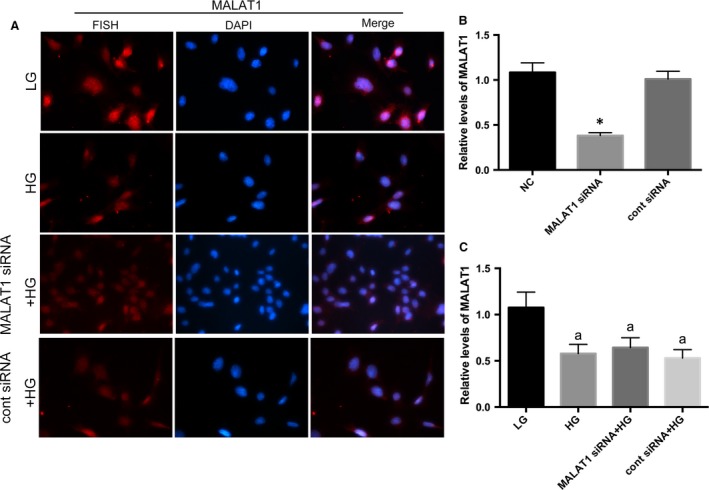
Determination of MALAT1 location mouse podocytes under culture. (A) By FISH, we observed a nuclear location of MALAT1 in cultured mouse podocytes. HG for 48 hrs significantly decreased MALAT1 staining, without resulting in any dislocation of MALAT1; however, MALAT1 siRNA transfection did not lead to a further extent of obliteration of MALAT1 staining, compared with podocytes without transfection. Podocytes under culture of LG or transfected with cont siRNA were the controls. (B) MALAT1 knock‐down efficiency was examined by real‐time PCR, which showed a significant abrogation of MALAT1 after transfection of MALAT1 siRNA for 48 hrs. Podocytes under normal conditions or transfected with cont siRNA were the controls. *P < 0.01 versus NC or cont siRNA. (C) Real‐time PCR analysis showed that HG treatment for 48 hrs resulted a remarkable reduction in MALAT1 levels; MALAT1 siRNA transfection did not lead to a further extent of obliteration of MALAT1 staining, compared with podocytes without transfection. Podocytes under culture of LG or transfected with cont siRNA were the controls. Values denote the mean ± S.D.; aP < 0.01 versus LG.