
Figure 3.
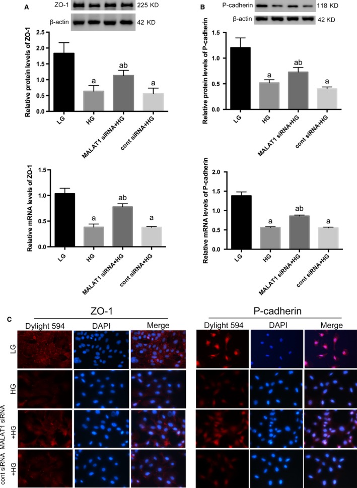
Determination of cellular markers of podocytes. (A,B) Western blot and real‐time PCR showed specific markers ZO‐1 and p‐cadherin were abundantly expressed in podocytes under LG conditions, both of which were significantly prohibited by HG on both protein and mRNA levels. And these abnormalities were abrogated by MALAT1 knock‐down. Podocytes under culture of LG or transfected with cont siRNA were the controls. Values denote the mean ± S.D.; aP < 0.01 versus LG,bP < 0.05 versus HG. (C) Immunofluorescence showed a 48‐hrs incubation with HG led to decreased staining of p‐cadherin and ZO‐1, in comparison with cells under LG conditions when ZO‐1 was intensely located at cell‐cell contacts and p‐cadherin highly expressed at cytoplasm and nuclei; these alterations were partially rectified by MALAT1 siRNA transfection, presenting early prohibition of MALAT1 was protective for HG‐treated podocytes. Magnification 400 × .