
Figure 5.
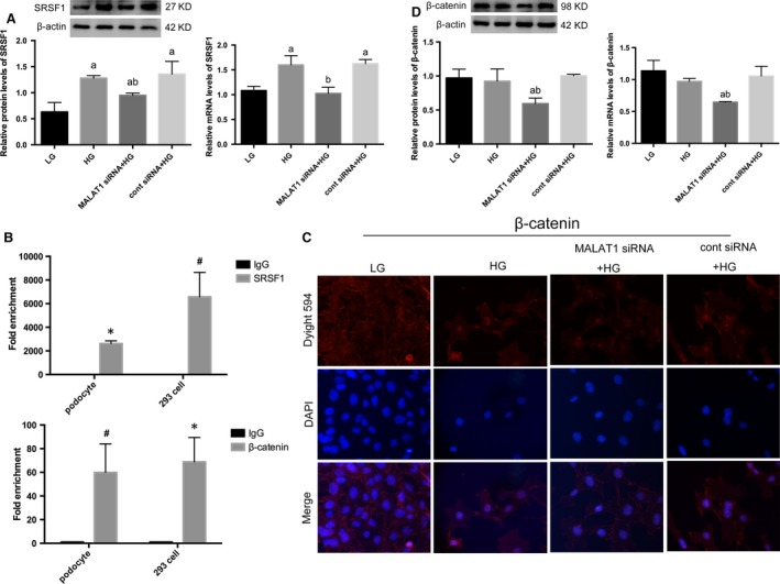
Effects of MALAT1 knock‐down on β‐catenin and SRSF1. (A) A 48 hrs of HG incubation was sufficient to result in an overexpression of SRSF1, compared with LG; this mutation was partially reversed by MALAT1 siRNA transfection. Podocytes under culture of LG or transfected with cont siRNA were the controls. Values denote the mean ± S.D.; aP < 0.01 versus LG,bP < 0.05 versus HG. (B) RIP analysis using anti‐SRSF1 or anti‐β‐catenin antibodies were performed either in cultured mouse podocytes with or without β‐catenin shRNA transfection, or in human 293 cells. A control IgG was used as the negative control for immunoprecipitation. Real‐time PCR was used to determine the RIP signals. Values denote the mean ± S.D.; *P < 0.01 versus IgG, # P < 0.05 versus IgG. (C,D) Immunofluorescence showed β‐catenin nuclear translocation was triggered by HG stimulation, although total protein levels were not significantly altered; MALAT1 knockdown significantly down‐regulated β‐catenin expression on both protein and mRNA levels, with diminished high glucose‐induced nuclear accumulation of β‐catenin. Podocytes under culture of LG or transfected with cont siRNA were the controls. Values denote the mean ± S.D.; aP < 0.01 versus LG,bP < 0.01 versus HG.