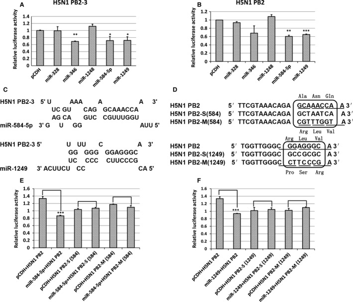
Figure 2.

Confirmation of miR‐584‐5p and miR‐1249 binding sites on the IAV PB2 gene. (A, B) The relative luciferase activity of PB2‐3 or PB2 in 293FT cells overexpressing various miRNAs. The experiments were carried out in triplicate and repeated twice. Because the relative luciferase activity was different in every repeat, however the inhibition effect was in general the same, so the relative luciferase activity of the pMIR‐REPORT vector was set as 1 to compare conveniently. Data are expressed as mean ± S.D., n = 6. All statistical analyses were carried out (with pCDH as the control) using the t‐test. *P < 0.05, **P < 0.01, ***P < 0.001. (C) The model of hybridization between miR‐584‐5p or miR‐1249 and pb2‐3 mRNA predicted using RNAHybrid software. (D) The synonymous (S) and missense mutant (M) nucleotide sequences of PB2 for the miR‐584‐5p and miR‐1249 binding sites. (E, F) The relative luciferase activity of PB2 in cells transfected with S/M mutant sequences of PB2 in 293FT cells overexpressing either miR‐584‐5p (E) or miR‐1249 (F) after infection with H5N1 IAV. The experiments were carried out in triplicate and repeated three times, and data are expressed as mean ± S.D., n = 3. All statistical analyses were carried out using the t‐test. ***P < 0.001.
