
Figure 1.
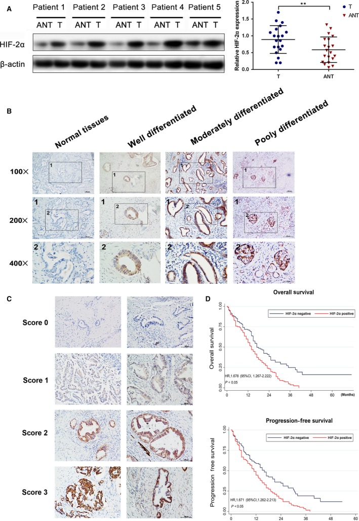
Up‐regulation of HIF‐2α correlated with poor prognosis in human PDAC . (A) Western blot analysis and qRT‐PCR analysis of HIF‐2α expression in PDAC tissues (T) and matched adjacent non‐tumour tissues (N). The mRNA and protein levels were normalized to β‐actin. **P < 0.01 (B) The images of immunohistochemistry for each differentiation degree in paraffin‐embedded PDAC and non‐tumours tissues. (C) The representative images for each score were showed. (D) The relationship between the expression of HIF‐2α and OS or PFS.