Figure 3.

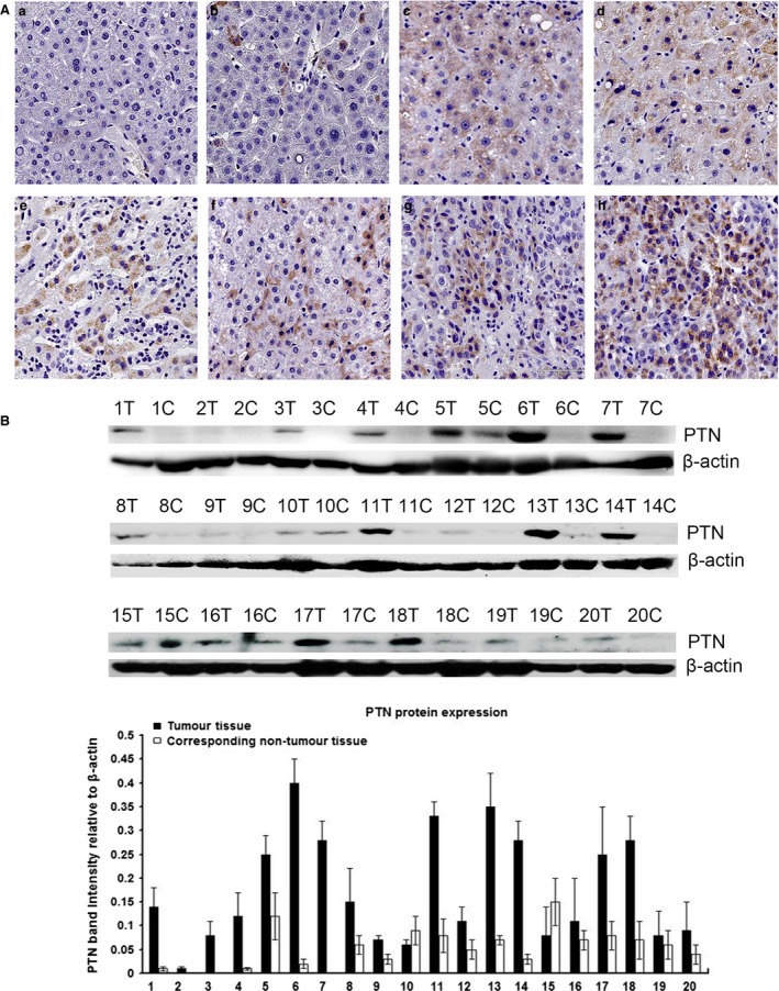
Analysis of the expression level of PTN in tissue. (A) PTN expression was much higher in HCC tissues than in non‐neoplastic and normal tissues. The IHC results showed that PTN was expressed in HSCs, hepatocytes and hepatoma cells, PTN protein in HCC was localized in the cytoplasm and the expression of PTN was elevated in some patients with steatosis (a: normal hepatic tissues; b: matched non‐neoplastic tissues; c and d: hepatitis tissues with partial steatosis; e and f: hepatic cirrhosis tissues; g and h: HCC tissues). (B) Western blot results showing that the expression of PTN was significantly higher in HCC tissues than in matched non‐neoplastic tissues.
