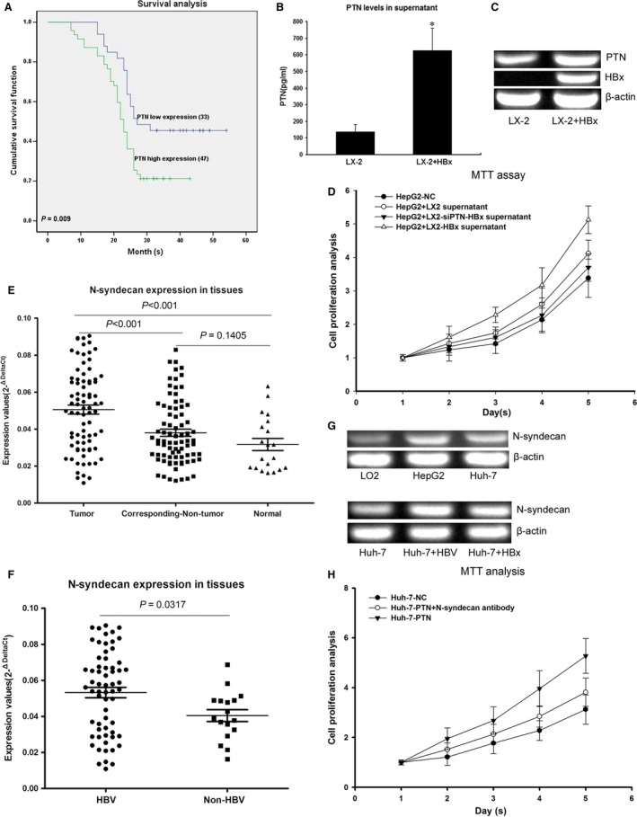
Figure 5.

HBx stimulates the growth of hepatocytes through PTN expression, and the PTN receptor N‐syndecan is highly expressed in HCC tissues. (A) Kaplan–Meier analysis showing that HCC patients with high PTN expression presented significantly shorter survival time than those with low PTN expression. (B) Comparative analysis of PTN levels in the supernatant of LX‐2‐HBx and control cells by ELISA. PTN levels were higher in LX‐2‐HBx than in controls. (C) RT‐PCR results showing that PTN mRNA levels were higher in LX‐2‐HBx than in controls. (D) HepG2 displayed the most rapid proliferation in the supernatant of LX‐2‐HBx. The expression of PTN was inhibited by RNAi in LX‐2‐HBx cells and cocultured with HepG2. This result showed that the proliferation of HepG2 cocultured with LX‐2‐HBx‐siPTN was reduced compared with the control. (E) Analysis of 80 paired tumour tissue samples, adjacent non‐tumour tissue samples and 20 normal tissue samples showed that the expression of N‐syndecan was increased in tumour tissues compared with non‐tumour and normal tissues. (F) HBV‐infected HCC patients showed higher expression levels of N‐syndecan compared with that in HBV‐negative HCC patients. (G) The expression of N‐syndecan in HepG2 and Huh‐7 was higher than that in LO2 normal hepatocytes, and the expression of N‐syndecan was up‐regulated in HBV‐infected cells and HBx‐transfected cells compared with control cells. (H) Antibody inhibition of N‐syndecan could impede hepatocyte proliferation induced by PTN. *P < 0.05
