Figure 7.

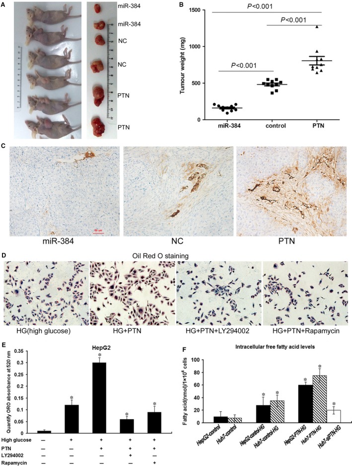
MiR‐384 and PTN affect tumour growth and angiogenesis in vivo, and PTN promotes lipogenesis. (A) Huh‐7 cells (5 × 10⁶) transfected with miR‐384, PTN and controls were implanted subcutaneously into the flank of nude mice. The mice were killed 45 days later, and tumour nodules were removed and photographed. (B) The tumour nodule weight in the PTN overexpression group was greater than that in the control group. The tumour nodule weight in the miR‐384 overexpression group was smaller than that in the control group. (C) Immunohistochemical staining with CD34 antibody was conducted to analyse the microvessel density in tumour nodules. The density of tumour blood vessels observed in PTN overexpression tumour nodules greatly exceeded that in the control group and the miR‐384 overexpression group. (D) The cells were stained with Oil Red O. Lipogenesis induced by high glucose was much higher in HepG2‐PTN than in HepG2‐NC. The lipogenesis induced by high glucose in HepG2‐PTN was inhibited by LY294002 and rapamycin. (E) ORO staining was quantified by measuring the absorbance at 520 nm. The lipid content of HepG2‐PTN cells was significantly higher than that of the parental control cells, and LY294002 and rapamycin could diminish lipogenesis. (F) The high sugar could promote free fatty acid synthesis. The intracellular free fatty acid levels in PTN‐transfected cells were higher than those in control cells. Down‐regulation of PTN expression weakened HG‐induced lipogenesis. *P < 0.05
