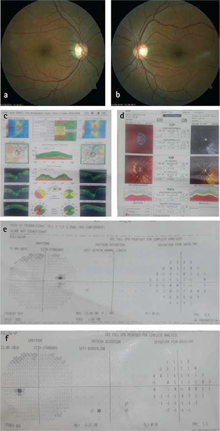
Figure 2. A patient with history of multiple sclerosis-associated retrobulbar optic neuritis. Optic disc examination, visual field test, and Heidelberg retinal tomography are within normal limits, while optical coherence tomography reveals retinal nerve fiber layer defect in the right eye.