
Figure 3.
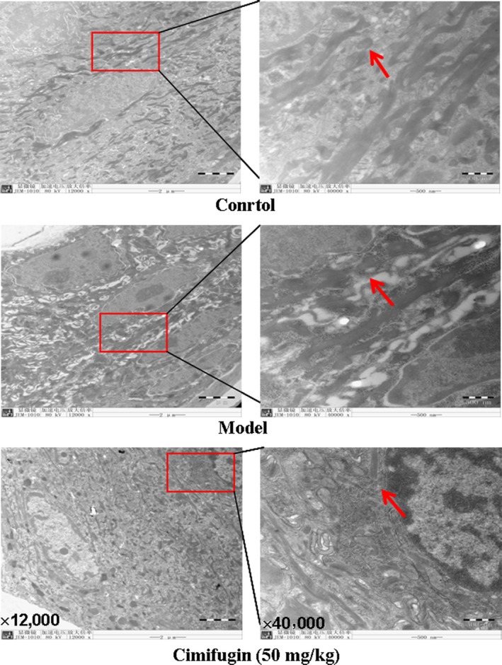
Effects of cimifugin on the ear skin epithelium in the initial stage of AD model. An electron microscopy observation has been performed on ears tissues of each group (n = 3, magnification: ×12,000; ×40,000). Arrowheads indicate the junction gap of epithelium. The data are representatives of three independent experiments.