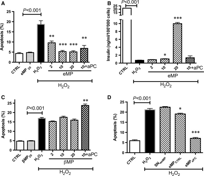
Figure 1.

Effect of aPC alone or of aPC‐generated microparticles on β‐cells submitted to oxidative stress. (A–B) β‐cells were pre‐treated by aPC (70nM, 4 hrs) or endothelial cell‐derived MP treated by aPC (eMPaPC, 6 hrs) before the 24 h‐H2O2 treatment. Apoptosis was assessed by hypodiploid DNA labelling (A, n = 4). Insulin secreted in supernatant was measured by ELISA (B, n = 4). (C) β‐cells cells were pre‐treated with β‐cells‐derived MP (ßMP) during 6 hrs before treatment by oxidative stress, in the absence or presence of 50 nM aPC (aPC n = 4). (D) β‐cells were treated by the supernatant of control untreated endothelial cells (SNwoMP) or by MP harvested from untreated resting endothelial cells (MPCTRL) during 6 hrs prior addition of H2O2.. Data expressed as mean ± S.E.M. (aPC, activated protein C; CTRL, untreated cells; eMP, microparticles isolated from aPC‐treated endothelial cells; βMP, microparticles from β‐cells treated by aPC; PhSer eq., Phosphatidylserine equivalent. *P < 0.05 versus H2O2; **P < 0.01 versus H2O2; ***P < 0.001 versus H2O2).
