Figure 1.

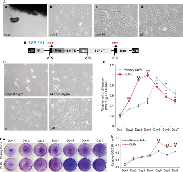
Immortalization of human cranial suture progenitor cells (iSuPs) derived from the patent suture tissues from craniosynostosis patients. (A) Primary suture progenitor cells (SuPs) were isolated from the freshly harvested cranial sutures and cultured in complete DMEM medium (a). Morphology of the recovered primary cells was recorded at day 9 (b) and day 14 (c) after plating, as well as at passage 3 (p3) (d). (B) Schematic representation of the reversible immortalization vector SSR #41. This retroviral vector contains the hygromycin and SV40 T antigen expression cassette flanked with FRT sites and can be removed by the Flippase (FLP) recombinase. (C) Establishment of iSuPs. The primary SuP cells were infected with packaged SSR #41 and selected in hygromycin‐containing medium for 5 days. Survived cells were observed at day 5 and day 14 post‐selection. The iSuPs were seeded at low density and passed consecutively for 3 (p3) or 20 passages (p20). Representative images are shown. (D) Cell proliferation evaluated by WST‐1 assay. The same number of primary SuPs (passage 2) and iSuPs was seeded at a low density. WST‐1 substrate was added to the cell culture and assessed for A450 nm readings at the indicated time‐points. Assays were performed in triplicate. ‘**’P < 0.001. (E) Cell proliferation assessed by crystal violet staining assay. The same number of primary SuPs and iSuPs was plated with a low density and fixed for crystal violet staining at the indicated time‐points (a). The stained cells were dissolved for OD reading and quantitatively determined at A590 nm (b). The assays were performed in three independent batches of experiments. Representative results are shown. ‘**’P < 0.001.
